Antibodies: The future is hybrid (Part 1)
Antibodies: the Future is Hybrid (Part 1)
Welcome to the first instalment of our three-part series exploring the ever-evolving world of scientific understanding. At Scientific Group, our curiosity drives us to stay at the forefront of innovation. It’s what makes us experts in crafting compelling stories that elucidate complex medical information. Stay tuned over the coming weeks for the next parts of this series, where we'll continue to explore a recent discovery that challenges our understanding of antibodies, and its implications on immunology.
To protect us, the immune system must recognize intruders in many different shapes and sizes. Antibodies are natural molecules that form an essential part of our ‘fluid-phase’ recognition system, also known as our humoral immunity.1 These protective proteins are named after their primary function: to bind tightly to all manner of foreign elements (from the German Antikörper: a substance raised against a foreign body 2).
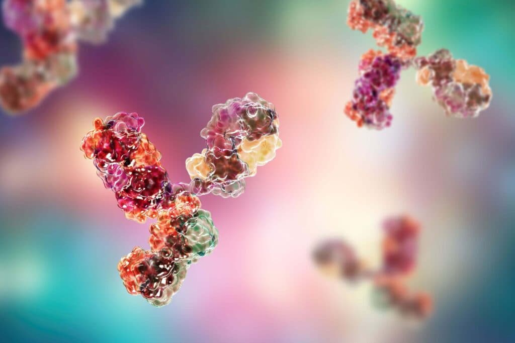
Antibodies are Y-shaped molecules with two ‘prongs’ at the top (variable regions) that bind their targets while the ‘base’ (constant region) assigns them different protective abilities1. Using different ‘bases’, antibodies fight invading forces in two main ways: by simply blocking foreign molecules when they attach or by coating pathogens and flagging them for destruction.1
Most antibodies have a relatively simple architecture, consisting just of two different polypeptides: a heavy chain and a light chain.3 For that reason, unique antibodies that bind to the same specific place on a target (i.e., monoclonal antibodies) are relatively simple to engineer and produce—a feature which has made them very popular for the treatment of human disease. In fact, as of July 2024, there are 168 different monoclonal antibody therapies either approved or in regulatory review across the European Union and the United States.4 Several new classes of therapeutic antibody-derived molecules have been developed, including fragments of whole antibodies (Fabs), antibodies that bind two or three different targets (bi/tri-specifics), and antibodies fused to other molecules (e.g., antibody-drug conjugates).3,5
Given that we understand antibodies well, and have studied them for over a century,6 one might expect there to be no more surprises in the world of antibody biology. Instead, a startling discovery has recently been made that challenges our current paradigm for antibody specificity.
The origin of antibody specificity The antibody repertoire diversity, or the number of unique antibodies in each individual, is estimated to be in the hundreds of billions.7 This staggering diversity is created through a combination of controlled genetic shuffling followed by a barrage of mutations in specific regions, processes called V(D)J recombination and somatic hypermutation, respectively (Figure 1). For a very long time, these two mechanisms were thought to be the only ways antibody diversity could be generated—until 2016, when a breakthrough came out of left field: from malaria research. Malaria is a disease in which antibodies play an important role. As the parasite invades our red blood cells, antibodies can either block the invasion or inhibit the function of parasite-derived proteins that are expressed on the red blood cell surface.8 ‘Broadly neutralizing’ antibodies (i.e., antibodies that would be effective against multiple types of malaria) are particularly effective at treating disease and can be generated naturally in one patient but used to fight malaria in others. In the hopes of discovering such therapeutically useful antibodies, the authors of this breakthrough study began sequencing antibodies from Kenyan malaria survivors.9 However, instead of the usual gene structure of a normal antibody, the investigators found a massive region of DNA inserted from a completely different part of the genome (Figure 1).9 This genetic ‘hitchhiker’ came from part of the DNA that encodes a receptor called LAIR1 which, strangely, had nothing to do with antibodies – or malaria! Further study revealed that the product of this genetic abnormality was a hybrid protein composed of the binding moiety of LAIR1 spliced onto the scaffold of a normal antibody (Figure 1)10. Finally, the fact that there were multiple hybrid antibodies, and that they could neutralize malaria growth cultures, suggested that this was a general mechanism of generating malaria-specific antibodies rather than a fluke.9
Figure 1. Comparison of a conventional antibody (left) and a hybrid antibody (right). In a conventional antibody, specificity is determined by the combinatorial diversity generated by V(D)J recombination. In a hybrid antibody, specificity is determined by the inserted sequence. A simple schematic of the genetic mechanism that generates each is depicted below.
*Light chain rearrangement not shown. Red arrows indicate mutations introduced to generate further diversity. Chr=chromosome; LAIR1=leukocyte-associated immunoglobulin-like receptor 1.
Pieces of the puzzle But what could the role of these hybrid anti-malaria antibodies be? In healthy individuals, LAIR1 normally binds collagen, a structural protein in connective tissue like cartilage, ligaments and tendons.11 So why does a collagen-binding receptor have properties that enables it to bind malaria-infected cells? Do hybrid antibodies exist outside of a malaria infection? And if so, why does the immune system need this third mechanism to generate binding diversity? If you’re curious to learn more—a value we embody at Scientific Group—then stay tuned for the answers to these questions and more in Part 2. Scientific Group has extensive experience of working in immunology, including dermatology, rheumatology, gastroenterology, and oncology. If you’d like to find out more, or are looking for expert agency support in this therapy area, please contact alejandro.potes@wearescientific.com
References
1. Murphy K, Weaver C. Janeway’s Immunobiology. 9th ed. Garland Science; 2017.
2. Lindenmann J. Origin of the terms “antibody” and “antigen.” Scand J Immunol. 1984;19(4):281-5.
3. Chiu ML et al. Antibody Structure and Function: The Basis for Engineering Therapeutics. Antibodies. 2019;8(4).
4. The Antibody Society. Antibody therapeutics approved or in regulatory review in the EU or US. Accessed July 8, 2024. https://www.antibodysociety.org/resources/approved-antibodies/
5. amsbio. Bispecific and Trispecific Antibodies. Accessed June 27, 2024. https://www.amsbio.com/bispecificandtrispecificantibodies
6. Behring E von, Kitasato S. Ueber das Zustandekommen der Diphtherie-Immunität und der Tetanus-Immunität bei Thieren. Deutsche Medizinische Wochenschrift. 1890;16:1113-14.
7. Glanville J et al. Precise determination of the diversity of a combinatorial antibody library gives insight into the human immunoglobulin repertoire. Proc Natl Acad Sci USA. 2009;106(48):20216-21.
8. Phillips MA et al. Malaria. Nat Rev Dis Primers. 2017;3(1):1-24.
9. Tan J et al. A LAIR1 insertion generates broadly reactive antibodies against malaria variant antigens. Nature. 2016;529(7584):105-9.
10. Hsieh FL, Higgins MK. The structure of a LAIR1-containing human antibody reveals a novel mechanism of antigen recognition. Elife. 2017;6.
11. Linsenmayer TF. Collagen. Cell Biol Extracell Matrix. 1991:7-44 (Published online).